Computer planned and guided implant surgery

The use of three-dimensional radiography and imaging (Computed Tomography {CT}, Magnetic Resonance Imaging {MRI}) has been used for more than four decades in medicine.
It has aided in increasing the accuracy of identification of vital anatomic structures and the pathologies associated within them.
This advanced technology has also prompted the development of protocols whereby surgical intervention can be planned on three-dimensional virtual computer animation or physical anatomic models. Today, computer-guided and robotic surgery in the most dangerous parts of the body such as the brain, spine and heart are routinely performed with great success and predictability.
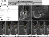
In dentistry, the introduction of 3-D radiography more than a decade ago has made it easier for the clinician to identify, study and plan a course of therapy to treat the area of disease or defect with increased precision (Fig. 1).
In addition, the introduction of office-based cone beam volumetric tomographic (CBVT) machines in 1999 came together with the advances in surgical planning software. This software comes either as a third party or as native to the image acquisition and viewing software included with the imaging hardware and has made implant therapy predictable and accurate (Fig. 2).

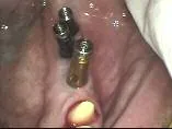
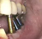
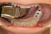
Traditional model-based surgical guides provide a reasonable estimation of the implant position for the prosthetic rehabilitation. The major limitations of these surgical guides was the surgery was often accomplished with flaps and the surgeon didn`t have an accurate estimation of the hard tissue present, especially the width, until the bone was exposed during surgery. This often led to surprises for both the surgeon and the patient, resulting in implants being placed that were under-engineered for the load or implants that later could not be restored esthetically, leading to compromised results (Figs. 3–7).


Computer-based implant planning and placement allows for creation of an exact replica of the jawbone on the computer screen, allowing visualization of all the vital structures such as nerves, sinuses, nasal floor, proximal teeth and concavities like the one below the mylohyoid ridge in the posterior mandible (Figs. 8a, b). Thus, practitioners can safely avoid these structures when planning and ultimately placing the implants using CAD/CAM generated surgical guides (Figs. 9–11).


With computer-guided placement of dental implants, there is no guesswork or surprises and most surgeries can be performed with a flapless technique (Figs. 12a–c). In case augmentation procedure has to take place, flaps can be reflected to access those sites and the implants provisionalized immediately (Figs. 13a–c). This conservative approach drastically diminishes postoperative pain, recuperation and healing time. The patient leaves the surgeon`s office esthetically restored and pleased with the ease at which such a complicated surgery was accomplished.


The guided surgical treatment is based on guided keyhole surgery that is minimally invasive. This reduces pain and swelling considerably for the patient compared to conventional treatment. This technique also reduces the number of appointments and chair time for the patient.
For many patients this means a considerable time and cost savings. The combination of immediate esthetic rehabilitation and function with temporary or final prosthesis ready at surgery radically shortens the overall treatment time and inconvenience to the patient. The computer-based surgical guides allow the implant surgeon to implement the planning with high precision and predictability. The use of a drilling template saves valuable chair time, and is a significant cost savings to the patient. The precision of a drilling template cannot be reproduced with the freehand method whether the task involves restorations of individual teeth or more extensive and elaborate implant planning.
Obtaining maximum certainty and safety through exact planning and precise implementation with a computer-based keyhole drilling template is both judicious and good patient care.
There are several implant planning software programs available, including: Galileos Implant from SiCat of Sirona, Procera from Nobel-Biocare and SimPlant from Materialise Dental, among others. All systems utilize a double scan technique for the evaluation of the implant site, planning the surgery and fabrication of the surgical guides.
When the patient consents to implant therapy, the restorative or surgical doctor first clinically evaluates the surgical area (Figs. 14a, b) and then refers the patient. If the clinician feels that there is adequate bone volume present to place the implant/implants in the proper position for acceptable esthetic and functional load, then an initial scan is not required.


Once the scan has been acquired, the preliminary implant planning can begin. The scan will aid in determining the amount of bone volume present to achieve primary implant stability, and the grafting required to augment the surgical site at the time of surgery. The implant planning can be easily shared with the entire implant team, including the patient, with the visual aid of the scan and computer. If it is determined from the scan that there is not enough bone volume to place the implant, then significant alteration in the existing anatomy is required prior to implant placement.
After implant planning, the patient is ready for a workup for the surgical guide fabrication. Study models are made (Fig. 15a) and the prosthetic laboratory will wax-up anatomically accurate teeth or a prosthesis as per the treatment plan. The technician will then convert the wax-up into an acrylic prosthetic replica of the final restoration made of a 25 percent barium sulfate and acrylic mixture and embed the replica in a clear retainer (Figs. 15b, c) attached to a scan template (radiographic or scan guide) (Figs. 15d, e) to be worn by the patient during a scan to be used for the final implant planning (Fig. 16a).


The scan template has fiduciary radiopaque markers that allow for accurate mounting of the stone model with the scan guide into the CAD/CAM milling machine that marks, drills and inserts the key hole sleeves into the scan guide, converting it into a surgical guide (Fig. 16b).

Following the simple process of marking the nerve canal and identifying vital proximal structures, the software offers the possibility of selecting from a wide variety of realistic implants from most implant manufacturers, and in a situation where a manufacturer has not provided the appropriate codes to the software company, the clinician can select a generic implant body and define the length as well as the apical and occlusal diameters.
The implant planning report along with the virtual implant placement, recorded on a CD-ROM, and the cast of the jaw where the implants are going to be placed are sent to the surgical guide manufacturer who will utilize CAD/CAM to fabricate a surgical guide with the appropriately sized sleeves embedded in the exact locations of the planned implants to accommodate the initial pilot drill of the implant system that will be used or with the sleeve-in-sleeve design for the entire surgery, including the insertion of the implant through the guide. The process is usually uneventful and the postoperative recovery is speedy.
By easily integrating the intuitively designed implant planning software into your implant practice, and utilizing a computer-generated surgical guide, the implant surgeon can achieve an easy, safe and predictable approach to implant planning and implant surgery.
The author wants to thank Dr. Jerome Kaufman, DDS (Pros.), of LeVisage Cosmetic & Implant Dentistry for his invaluable input and contribution toward the development of this article.
ImplantTribune May-2009 Computer Guided Implant Surgery and PRP_BMA.pdf